Tag Archives: best radiologist

Cancer patients want more information about medical imaging risk

Radiology_Radiation TherapyA substantial gap exists between patient expectations and current practices for providing information about medical imaging tests that use radiation, according to a new study published online in the journal Radiology. Researchers said the findings highlight a need for better communication as medicine enters an era of patient-centered care.In recent years, there have been numerous reports in the media about potential risks of tests that use ionizing radiation. However, benefit-risk discussions about ionizing radiation from medical imaging are rare and seldom initiated by clinicians.For the new study, researchers from Memorial Sloan Kettering Cancer Center (MSKCC) in New York City analyzed over nine hours of transcribed conversations with 30 people who had undergone medical imaging exams to determine their understanding of the benefits and risks associated with various medical imaging procedures and their expectations regarding communication of those benefits and risks.

Read the rest of the article at http://www.medicalnewstoday.com/releases/291385.php.

Cherenkov Effect improves radiation therapy for patients with cancer

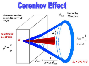
Radiation TherapyThe characteristic blue glow from a nuclear reactor is present in radiation therapy, too. Investigators from Dartmouth’s Norris Cotton Cancer Center, led by Brian W. Pogue, PhD, and PhD candidates Adam K. Glaser and Rongxiao Zhang, published in Physics in Medicine and Biology how the complex parts of the blue light known as the Cherenkov Effect can be measured and used in dosimetry to make therapies safer and more effective.”The beauty of using the light from the Cherenkov Effect for dosimetry is that it’s the only current method that can reveal dosimetric information completely non-invasively in water or tissue,” said Glaser.

Read the rest of the article at http://www.medicalnewstoday.com/releases/290027.php.

Sunlight continues to damage skin in the dark

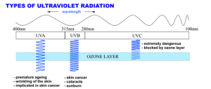
Dermatology_Radiology_OncologyMuch of the damage that ultraviolet radiation (UV) does to skin occurs hours after sun exposure, a team of Yale-led researchers concluded in a study that was published online by the journal Science.Exposure to UV light from the sun or from tanning beds can damage the DNA in melanocytes, the cells that make the melanin that gives skin its color. This damage is a major cause of skin cancer, the most common form of cancer in the United States. In the past, experts believed that melanin protected the skin by blocking harmful UV light. But there was also evidence from studies suggesting that melanin was associated with skin cell damage.In the current study, Douglas E. Brash, clinical professor of therapeutic radiology and dermatology at Yale School of Medical, and his co-authors first exposed mouse and human melanocyte cells to radiation from a UV lamp. The radiation caused a type of DNA damage known as a cyclobutane dimer (CPD), in which two DNA “letters” attach and bend the DNA, preventing the information it contains from being read correctly. To the researchers’ surprise, the melanocytes not only generated CPDs immediately but continued to do so hours after UV exposure ended. Cells without melanin generated CPDs only during the UV exposure.

Read the rest of the article at  http://www.medicalnewstoday.com/releases/289728.php.

Human stem cells repair damage caused by radiation therapy for brain cancer in rats

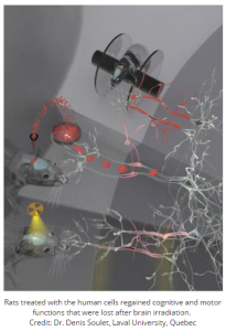
Nuclear Medicine_RadiologyFor patients with brain cancer, radiation is a powerful and potentially life-saving treatment, but it can also cause considerable and even permanent injury to the brain. Now, through preclinical experiments conducted in rats, Memorial Sloan Kettering Cancer Center researchers have developed a method to turn human stem cells into cells that are instructed to repair damage in the brain. Rats treated with the human cells regained cognitive and motor functions that were lost after brain irradiation. The findings are reported in the February 5 issue of the journal Cell Stem Cell.During radiation therapy for brain cancer, progenitor cells that later mature to produce the protective myelin coating around neurons are lost or significantly depleted, and there is no treatment available to restore them. These myelinating cells–called oligodendrocytes–are critical for shielding and repairing the brain’s neurons throughout life.A team led by neurosurgeon Viviane Tabar, MD, and research associate Jinghua Piao, PhD, of the Memorial Sloan Kettering Cancer Center in New York City, wondered whether stem cells could be coaxed to replace these lost oligodendrocyte progenitor cells. They found that this could be achieved by growing stem cells–either human embryonic stem cells or induced pluripotent stem cells derived from skin biopsies–in the presence of certain growth factors and other molecules.

Read the rest of the article at  http://www.medicalnewstoday.com/releases/289083.php.

Majority of primary care physicians find that medical imaging improves patient care

RadiologyAccording to a study published online in the Journal of the American College of Radiology (JACR), large majorities of primary care physicians believe that advanced medical imaging, such as computed tomography (CT), magnetic resonance imaging (MRI) and positron emission tomography (PET), provides considerable value to patient care.

A national survey of 500 primary care physicians was conducted using an online self-administered questionnaire. Study results showed that primary care physicians overwhelmingly indicated that advanced imaging:  For more information read here http://www.medicalnewstoday.com/releases/288560.php

Patient-centred approach to recording side effects of radiotherapy

Radiology

For lung cancer that cannot be removed with surgery, radiotherapy is the primary treatment option. However, it is associated with a range of side effects, including fatigue and inflammation of the oesophagus and lungs.

Current methods to record treatment-related toxicities rely on assessment by health care professionals. Now a team from The University of Manchester and The Christie NHS Foundation Trust – both part of the Manchester Cancer Research Centre – has explored the use of patient-reported outcomes to improve the recording of side effects for lung cancer patients.

Dr Corinne Faivre-Finn, a researcher in The University of Manchester’s Institute of Cancer Sciences and a consultant based at The Christie NHS Foundation Trust, who led the research, said: “Such patient-reported outcome tools have been mainly evaluated for use with chemotherapy treatments. We wanted to assess their feasibility and relevance in lung cancer patients undergoing radiotherapy.”

The group looked at the agreement between side effects as reported by doctors and the patients themselves. They also evaluated the relationship between reported toxicities and quality-of-life measures, relating to aspects such as tiredness, anxiety and shortness of breath.

Patients were asked to fill in questionnaires covering both side effects and quality of life at three time points: before treatment, at the end of radiotherapy and at later follow up. The consultants answered questions at identical time points covering the same common radiotherapy-related toxicities for each patient.

The study found that there was strongest agreement between the patient’s scoring of side effects and measures relating to their quality of life. Toxicities as recorded by the clinicians appeared to underestimate their severity.

“This was the first study in Europe to explore such a patient-centred approach to recording side effects. Incorporating this method into cancer care could allow us to detect and manage serious effects earlier. It could also improve patient-doctor relationships and help doctors better understand the full impact of treatment on patients,” added Dr Faivre-Finn.

Adapted by MNT from original media release


http://www.medicalnewstoday.com/releases/287751.php

Real-time radiation monitor can reduce radiation exposure for medical workers

Radiology

It’s a sound that saves. A “real-time” radiation monitor that alerts by beeping in response to radiation exposure during cardiac-catheterization procedures significantly reduces the amount of exposure that medical workers receive, UT Southwestern Medical Center researchers found.

In a randomized study, the researchers divided 505 patients undergoing either diagnostic coronary angiography or percutaneous coronary intervention, such as stent placement, into two groups. In half the procedures, medical workers used the current gold standard for radiation monitoring, which is a “dosimetry” badge that is worn by a medical worker for a month and then sent off for the cumulative radiation dose to be read. In the other half, medical workers wore a device called Bleeper Sv, which beeps approximately once every 15 minutes in response to low background radiation, and beeps once every 20 seconds when exposure is higher, or continuously, if it is very high.

In settings where the medical workers wore the device that gives the real-time auditory feedback, radiation exposure was consistently decreased by approximately one-third.

“Radiation is invisible,” said Dr. Emmanouil Brilakis, Associate Professor of Internal Medicine at UT Southwestern and senior author of the paper. “Use of a radiation detection device can provide real-time ‘visualization’ of radiation exposure, enabling operators to take actions to reduce radiation exposure.”

Among actions that medical workers can take to reduce radiation exposure are reducing the frame rate (the number of X-ray images taken per second to create a “movie” of the coronary arteries), decreasing fluoroscopy time, repositioning the patient, repositioning the medical worker, adjusting the position of the radiation shield, and using additional shielding.

“Using devices that provide real-time radiation-exposure feedback can help the operator adopt safer radiation practices,” said Dr. Brilakis, who is also Director of the Cardiac Catheterization Laboratories at the VA North Texas Health Care System. “In our study, this was achieved in a real-life setting among unselected patients using a low-cost device that can be used with any X-ray system.”

Physicians and other medical workers on cardiac-catheterization teams will likely participate in hundreds of procedures a year. The dose limit for occupational exposure is 20 mSv per year for five years, but no dose is safe and all doses are considered to contribute to cancer risk.

“It has been shown that people who are chronically exposed to radiation in cardiac catheterization labs are more likely to develop left-sided brain tumors,” said Dr. Brilakis. “The reduction in operator exposure observed in our study is likely to translate into a decreased risk for long-term adverse clinical events.”

The results of the RadiCure study appear in the Dec. 16 issue of Circulation: Cardiovascular Interventions.

Adapted by MNT from original media release

http://www.medicalnewstoday.com/releases/287142.php


Children being given unnecessary chest X-rays

Nuclear Medicine

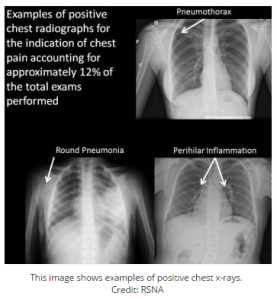
Researchers at Mayo Clinic found that some children are receiving chest X-rays that may be unnecessary and offer no clinical benefit to the patient, according to a study presented at the annual meeting of the Radiological Society of North America (RSNA).

“Chest X-rays can be a valuable exam when ordered for the correct indications,” said Ann Packard, M.D., radiologist at the Mayo Clinic in Rochester, Minn. “However, there are several indications where pediatric chest X-rays offer no benefit and likely should not be performed to decrease radiation dose and cost.”

Dr. Packard and co-author, Kristen B. Thomas, M.D., head of the pediatric division at Mayo Clinic and assistant professor of radiology at Mayo Medical School, reviewed data from 719 pediatric chest X-ray exams ordered between 2008 and 2014 in Mayo Clinic’s inpatient, outpatient and emergency room settings. The patients undergoing the exams ranged in age from newborn to 17 years old.

Of the 719 X-ray exams, 377 exams were ordered for chest pain, 98 indicated syncope (fainting) or presyncope, 21 indicated spells (a general feeling of being unwell or under distress), 37 indicated postural orthostatic hypotension (POTS)–a condition in whichblood pressure drops suddenly when the individual stands up from sitting or lying down, 185 indicated dizziness, and one exam indicated cyclical vomiting. Eighty-two of the 719 exams were excluded due to congenital or other known heart disease, and other causes.

The researchers found that in approximately 88 percent of the remaining 637 patients, the exam did not alter clinical treatment.

None of the patients who underwent X-rays for indications including syncope, spells, POTS, dizziness or cyclical vomiting had any finding that affected treatment. Thirty-nine of the 330 non-excluded X-rays for chest pain were positive for pneumonia, bronchial inflammation, trauma, or other conditions.

“Approximately 12 percent of the chest X-rays for chest pain were positive and included respiratory symptoms such as cough, fever or trauma,” Dr. Packard said. “There were no positive findings in any chest X-ray for syncope, dizziness, spells, cyclical vomiting or POTS for the past five years, even in our tertiary care center with referrals for rare diseases or unusual presentations.”

Optimizing radiation exposure and cost effectiveness are important topics in today’s healthcare environment, particularly in a pediatric population, Dr. Packard noted.

“This study addresses both of these issues, which is important not only for physicians but also for young patients and their parents,” she said. “I would like this research to help guide clinicians and deter them from ordering unnecessary exams which offer no clinical benefit to the patient.”

Adapted by MNT from original media release

http://www.medicalnewstoday.com/releases/286442.php


Surgery plus chemoradiation linked to improved survival rates for gastric cancer patients

Gastroenterology_Radiology

Patients who receive chemotherapy and radiation after surgery for gastric cancer appear to have better survival rates than those who had surgery followed by only chemotherapy, according to results of a look-back study of more than 500 people by Johns Hopkins scientists.

The combination of post-operative chemotherapy and radiation, or chemoradiation, resulted in survival for an average of 46.7 months after treatment, compared to 20.9 months among those who received chemotherapy only after surgery. Five years after treatment, 46.9 percent of chemoradiation-plus-surgery patients were still living, while 24.9 percent of chemotherapy-plus-surgery patients survived.

Chemoradiation also improved patients’ recurrence-free survival, to an average of 35.6 months, compared with 16.6 months among those who received chemotherapy alone.

The addition of radiation especially improved survival rates among gastric cancer patients whose cancers had spread to lymph nodes in the immediate region of the stomach, says Timothy Pawlik, M.D., M.P.H., Ph.D., professor of surgery at the Johns Hopkins University School of Medicine and member of the Johns Hopkins Kimmel Cancer Center.

Pawlik says the role of radiation therapy in treating gastric cancer has been understudied, “but these data would suggest that radiation therapy would benefit patients, in particular those patients who had disease that has spread to lymph nodes.”

Although rates of gastric cancer have dropped in recent years in the United States, it is still the fourth most common cancer and the second-leading cause of cancer deaths worldwide.

The study results, described in the Annals of Surgical Oncology, is a so-called retrospective one, looking at the records of 505 gastric cancer patients treated between 2000 and 2012. Retrospective studies come with a certain amount of limitation and bias, Pawlik says, because researchers can only analyze how therapy was carried out and can’t control which patients received which treatments.

Pawlik and his colleagues attempted to remove some of that bias in their study with a statistical fix. Using information on the patients’ ages, tumor sizes and other factors collected from the multi-institutional U.S. Gastric Cancer Collaborative database, the researchers were able to build a score that measured how likely it would be for a patient to receive chemoradiation along with surgery. Within groups of patients with similar scores, they were able to compare the outcomes between those who did get chemoradiation and those who had chemotherapy only.

This “fancy matching” helps get rid of some but not all of the bias in a retrospective study, says Pawlik. “It is possible that patients who got radiation therapy were younger, healthier, and could more easily tolerate radiation therapy, which factored into longer survival.”

Chemoradiation therapy also has improved over the past decade, making it an important tool to consider when treating gastric cancer, says Joseph Herman, M.D., director of clinical research in the Department of Radiation Oncology and Molecular Radiation Sciences at Johns Hopkins. “Some patients will benefit more from chemoradiation, and these large studies can give us more insight into who these patients might be.”

Other researchers involved in the study include Aslam Ejaz, Gaya Spolverato and Yuhree Kim of the Johns Hopkins University School of Medicine; Malcolm H. Squires and Shishir K. Maithel of Emory University; George Poultsides and David J. Worhunskyof Stanford University; Ryan Fields and Linda X. Jin of Washington University in St. Louis; Mark Bloomston, Carl Schmidt and Neil Saunders of The Ohio State University; Sharon M. Weber, Alexandra W. Acher and Clifford S. Cho of the University of Wisconsin; and Konstantinos Votanopoulos and Douglas Swords of Wake Forest University.

Ejaz was supported in part by the Eleanor B. Pillsbury Foundation for Surgical Research


Interstitial lung disease is a significant risk factor for lung inflammation following stereotactic body radiation therapy for lung cancer

Radiology

Pretreatment interstitial lung disease (ILD) is a significant risk factor for developing symptomatic and severe radiation pneumonitis in stage I non-small cell lung cancer (NSCLC) patients treated with stereotactic body radiation therapy (SBRT) alone.

ILD is a group of diseases that cause scarring and stiffing of the tissue and space around the air sacs in the lungs, which results in diminished gas exchange. The incidence of ILD among lung cancer patients is higher than in the general population as tobacco smoking is a common risk factor for both. Some lung cancer patients with ILD may not be considered good candidates for surgical therapy. SBRT uses sophisticated techniques to deliver a targeted and focused radiation dose to a tumor in order to stop the growth locally with limited damage to surrounding healthy tissue. SBRT is considered an acceptable therapy choice for early-stage NSCLC patients who are not good candidates for or decline surgery.

In order to determine the optimal treatment for early-stage lung cancer patients with ILD, researchers at Kyoto University in Japan examined the incidence of radiation pneumonitis and the clinical outcomes in 157 patients who underwent SBRT alone for stage I NSCLC.

Results published in the Journal of Thoracic Oncology, the official journal of the International Association for the Study of Lung Cancer (IASLC), show that of the 157 patients who received SBRT for lung cancer therapy, 20 were identified as having pretreatment ILD. The presence of ILD was a significant risk factor for both symptomatic and severe radiation pneumonitis and the cumulative incidence of radiation pneumonitis increased significantly with worse ILD. Overall survival trended to be shorter in the ILD positive population but this was not statistically significant and may be accountable to the ILD itself. There were no differences in disease progression or local progression rates in patients with ILD versus those patients without.

“Our results suggest that the impact of ILD on radiation pneumonitis depends on the preexisting severity of the ILD findings and clinicians should be cautious when considering SBRT for those with significant ILD findings”, say the authors. “However, other than radiation pneumonitis, life-threatening complications after SBRT are rare. Thus, if the severity of ILD and the risk of radiation pneumonitis are carefully evaluated, SBRT is a curative-intent treatment option for those with early-stage NSCLC and pretreatment ILD.”

http://www.medicalnewstoday.com/releases/285268.php


Picture courtesy of www.nature.com